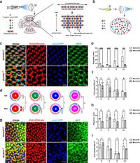
In a groundbreaking study, researchers have uncovered the critical role of Down Syndrome Cell Adhesion Molecule b (Dscamb) in shaping the mosaic structure of cone photoreceptors in zebrafish retinas. The study reveals that Dscamb is essential for maintaining proper spacing between different types of cone photoreceptors, particularly red cones, and its absence leads to significant disruption in their lattice-like arrangement.
The retina, crucial for vision, relies on the proper arrangement of cone cells, which are responsible for color detection. In zebrafish, these cones come in four types: red, green, blue, and ultraviolet, forming a regular mosaic pattern that allows for efficient photoreception. However, until now, the mechanisms governing this arrangement were poorly understood.
Utilizing CRISPR/Cas9 genome editing technology, researchers generated Dscamb mutant zebrafish. They discovered that in these mutants, the regular mosaic pattern of cones was compromised, especially noted through the clustering of red cones. During normal retinal development, red cones utilize filopodia—tiny, finger-like projections—to extend towards neighboring cones, engaging in a homotypic recognition process essential for maintaining order. In Dscamb mutants, the expected retraction of these filopodia was absent, causing red cones to cluster unnaturally.
Interestingly, while the disruption was most notable among the red cones, the study found similar behaviors in other cone types, although to a lesser degree. The robust methodology employed live imaging and focused on the dynamic interactions between photoreceptors as they matured. By observing these interactions, the researchers demonstrated how proper Dscamb function allows red cones to recognize and retract from other red cone apical surfaces, maintaining their spacing through a process identified as contact-dependent self-avoidance.
Cones form tight associations, yet their development must also accommodate differing types to optimize functionality in the retina. The importance of Dscamb, as evidenced in this study, highlights the intricate balance between homotypic and heterotypic interactions within retinal cellular development. The findings are significant, as they not only contribute to understanding the basic biology of cone arrangement in fish but may also shed light on visual system disorders in humans.
The research was conducted at a laboratory focused on developmental biology, and the findings were published recently, indicating a promising trajectory for future studies. The mechanisms explored could lead to insights into other molecules that assist in maintaining structural integrity in the retinas of various species. Further investigations may focus on other adhesion molecules that contribute to cone spacing and overall retinal integrity, potentially identifying new therapeutic targets for retinal degenerative diseases.
This study underscores the vital role that cell adhesion molecules play not just in neural spacing techniques but also in understanding complex visual systems and their evolution across species. As scientists continue to unravel the complexities of retinal development, the findings regarding Dscamb will undoubtedly guide future research into the fascinating world of cellular organization and its implications for vision.