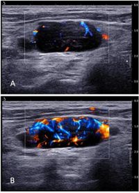
A recent study has shown that advanced ultrasonographic vascular imaging techniques can effectively differentiate between superficial invasive lymphomas and indolent lymphomas, which is crucial for improving patient diagnosis and treatment. The study, which included 82 lymphoma patients, employed multiple ultrasound methods—color Doppler flow imaging (CDFI), angio plus ultrasound imaging (AngioPLUS™), and contrast-enhanced ultrasound (CEUS)—to characterize lymph node blood flow and assess the aggressiveness of the cancers.
Numerous studies emphasize that lymphomas can be broadly classified as either invasive or indolent, a distinction that has significant implications for patient management. This classification is based on the lymphomas' proliferation rates and clinical courses. Invasive lymphomas, such as diffuse large B-cell lymphoma (DLBCL), pose a greater challenge due to their aggressive nature, while indolent lymphomas, like follicular lymphoma (FL), progress more slowly.
Led by researchers Lin Li and Wenjuan Lu, the study sought to assess whether the characterized blood flow in lymph nodes could reliably indicate the type of lymphoma present. Participants were all adults aged 18 and over, with histopathologically confirmed lymphoma types and superficial lymph nodes presenting high uptake on PET/CT scans. Over a timeline from September 2020 to November 2022, researchers employed various imaging techniques to capture intricate details about blood flow patterns.
According to the research findings, significant differences were identified across the ultrasound techniques used. For instance, CDFI allowed the classification of lymph nodes through a resistance index (RI), with invasive lymphomas showing a significantly higher RI than indolent lymphomas (0.66 ± 0.09 versus 0.56 ± 0.07, p < 0.001). Furthermore, AngioPLUS™ demonstrated superior blood flow performance and diagnostic sensitivity when compared to CDFI.
In contrast-enhanced ultrasound, substantial differences in necrosis and arrival time (ATM)—critical indicators of aggression—were noted between the two lymphoma types (p = 0.026 and p = 0.043, respectively). Ultimately, the combination of CDFI and CEUS techniques exhibited the highest diagnostic sensitivity of 98.1%. This remarkable finding points toward ultrasonographic vascular imaging's potential as a non-invasive, cost-effective diagnostic tool that could supplement traditional biopsy methods.
The diagnostic efficacy was further reinforced by excellent interobserver agreement for qualitative parameters assessed by the two radiologists involved in the study. The comprehensive data likely will lead to improved decision-making by clinicians when dealing with complex lymphoma cases.
The broader significance of this study cannot be overstated. Currently, invasive procedures such as lymph node biopsies are the gold standard for diagnosis. However, those methods can be risky, particularly for patients with significant comorbidities or those who are sensitive to general anesthesia. Ultrasonographic vascular imaging provides a non-invasive alternative that could reduce risks and improve patient outcomes. Given these advantages, the authors recommend its application in routine diagnostic pathways.
In summary, this research opens a promising pathway for more effective lymphoma diagnosis and treatment. The findings underline the pivotal role of ultrasonographic vascular techniques, which actively visualize the underlying pathophysiology of lymphomas. As clinicians continue exploring these techniques, the hope is that they will significantly advance lymphoma patient care and treatment strategies.
As with any clinical research, this study has some limitations: the sample size was relatively modest, and there was some selection bias since only superficial lymph nodes were examined. Future studies with larger sample sizes and diverse anatomical locations are necessary to further solidify these findings and potentially transform diagnostic protocols in lymphoma care.
The research has been positively received within the medical community and is expected to encourage further exploration into non-invasive imaging techniques as critical components of lymphoma diagnosis and treatment plans.