A new study utilizing 3D imaging technology reveals intricate details of root canal structures in maxillary premolars, which can enhance the success rates of root canal therapy.
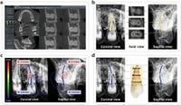
The success of root canal therapy (RCT) is heavily dependent on a comprehensive understanding of the root canal system's complexities. Traditional two-dimensional (2D) imaging methods, such as panoramic and periapical radiographs, often provide limited information, leading to challenges in accurately treating root canals. A recent study conducted at Yonsei University Dental Hospital aimed to address these limitations by employing advanced cone-beam computed tomography (CBCT) alongside specialized 3D analysis tools.
The research involved 400 maxillary premolars, with data collected from CBCT images between January 2020 and December 2022. Researchers connected the center points of the root canals to create virtual 3D models that allowed for more accurate assessments of canal morphology and curvature than previously possible. Remarkably, the study found that 99.5% of the first premolars and 88.0% of second premolars displayed double canals, with buccal and palatal branching.
Using the 3D analysis tool, researchers calculated the curvature of these canals, identifying the most curved points (MCPs) in this intricate network. The mean radius value of the MCPs was discovered to be 1.50 mm for first premolars and 1.58 mm for second premolars. The findings indicate that most MCPs were located in the apical region; however, a significant percentage, ranging from 12.1% to 35.2%, were identified to be in the middle or coronal parts.
As noted by the authors, "3D analysis tools on CBCT provide accurate information on the root canal system including the canal curvature, thereby increasing the success rate of RCT." This statement underscores the potential of this technology to reshape how dental professionals approach root canal therapies. By employing 3D visualization, clinicians can anticipate variations in canal shape and curvature, which are critical for effective treatment.
The methodology used in this study reflects a notable advancement in endodontic care. CBCT imaging, known for its ability to convey complex anatomical structures in three dimensions, has already been adopted across several areas of dentistry, but its application in endodontics has been limited until recently due to concerns over radiation exposure. However, with the advent of low-dose protocols, these concerns are being mitigated, paving the way for broader clinical applications.
In analyzing the root canal morphology, researchers also classified the canal types and examined the curvature at different locations within the root canal. The use of the 3D navigation software allowed for an intuitive analysis of curvature, facilitating the identification of critical anatomical features that might be missed through conventional imaging. The results indicated that severe curvature was present in all canals studied, with all maximum radius values being recorded below 4 mm, which is indicative of severe curvature.
Given that conventional 2D imaging can obscure essential details, the utilization of CBCT and 3D visualization tools marks a significant leap towards improving RCT outcomes. Utilizing this technology has previously led to changes in endodontic treatment plans in a notable number of cases, hinting at its growing importance in routine dental practice.
Ultimately, the findings from this study advocate for a paradigm shift in how dental professionals visualize and treat root canal systems, especially in maxillary premolars known for their anatomical complexity. By integrating 3D technology into clinical workflows, practitioners could enhance diagnostic precision and treatment effectiveness.
In conclusion, the research presents compelling evidence that 3D analysis provides not only detailed visualization but also crucial quantitative data on canal curvature that 2D methods simply cannot deliver. With better insights into the anatomy of root canals, dental professionals are equipped to make informed decisions that could ultimately improve patient outcomes in root canal therapy.