The complexity of human emotion regulation and how we navigate future threats is a crucial area of psychological research. A recent study using advanced brain imaging techniques has shed light on how individuals suppress thoughts of potential future threats and the resulting neural activity associated with this suppression. This study involved 65 participants who underwent an MRI scan while engaging in tasks of thought suppression and threat imagination. Notably, the research identified key brain regions actively involved during the suppression of anxiety-inducing thoughts.
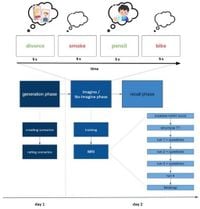
The study employed a well-established paradigm entitled the Imagine/No-Imagine Task (INI), focusing on how participants managed distressing scenarios related to their fears. Participants were asked to imagine fear-provoking events while simultaneously working to suppress these thoughts, creating a layered exploration of emotional regulation. As highlighted in the study, when participants engaged in suppression, significant activation was noted in regions such as the inferior frontal gyrus (IFG), middle frontal gyrus (MFG), superior parietal lobule (SPL), and superior occipital sulcus (SOS). These areas are believed to play pivotal roles in cognitive control and emotion regulation.
Conversely, when participants were instructed to imagine future threats, activation shifted dramatically to the bilateral posterior cingulate cortex (PCC) and the ventromedial prefrontal cortex (vmPFC). This finding underscores a fascinating neural shift that occurs depending on whether individuals are suppressing thoughts or imagining potential threats. The PCC, associated with introspection and self-awareness, coupled with the vmPFC's role in emotional regulation, signifies how our brains dynamically transition in response to varying emotional tasks.
To examine the network dynamics further, the researchers employed dynamic causal modeling (DCM) alongside group iterative multiple model estimations (GIMME). DCM unveiled that during suppression, strong connectivity links were observed from the MFG to critical regulatory areas, such as the vmPFC and the PCC. This neural architecture suggests a sophisticated interplay of brain pathways dedicated to managing and modulating emotional responses to anticipated threats.
The findings also demonstrated considerable variability in individual patterns of connectivity, suggesting the presence of distinct biotypes or profiles of emotional regulation strategies. This individual variability highlights the complex nature of mental health and the differing cognitive control mechanisms employed by individuals when confronting anxiety-inducing scenarios.
When analyzing participants' recall of suppressed threats, the study showed no significant differences in the recall accuracy of suppressed versus imagined threats, which poses interesting questions about the efficacy of thought suppression as an emotional regulation strategy. Despite previous findings suggesting diminished recall in suppressed scenarios, the current study did not replicate these results, warranting further investigation into the underlying dynamics of memory and suppression.
In conclusion, this study elevates our understanding of the neurological underpinnings associated with suppressing and imagining threats. By identifying specific brain regions and the associated networks activated during these emotional tasks, we can further explore potential clinical applications. Future research could aim to enhance suppression training through neuromodulation techniques, potentially improving mental well-being and customizing treatments for anxiety-related disorders.