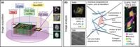
In a groundbreaking study published in Scientific Reports, researchers have developed a multimodal imaging workflow that significantly enhances the analysis of three-dimensional cell cultures, a pivotal aspect in biomedical research. This innovative approach combines fluorescence microscopy with advanced techniques including vibrational and second harmonic generation microscopy, secondary ion mass spectrometry imaging, and transmission electron microscopy. The integration of these methods allows for an in-depth characterization of cell-seeded scaffolds, essential for applications ranging from regenerative medicine to drug screening.
Conventional two-dimensional (2D) cell cultures often fall short of replicating the complex microenvironment found in living organisms, which can lead to misleading results in experimental studies. To remedy this, the scientific community has shifted towards using three-dimensional (3D) cell culture models. These models offer a more accurate representation of cellular interactions and physiological conditions. By understanding the cellular responses in these intricate environments, researchers can better evaluate therapeutic efficacy and toxicity, ultimately advancing personalized medicine.
The research team set out to investigate the structural, chemical, and morphological properties of human dermal fibroblasts (HDFs) seeded in type I collagen scaffolds. The study utilized a series of advanced imaging techniques to correlate multiple parameters, including cell distribution, cytoskeletal morphology, scaffold fiber organization, and biomolecular composition. Using cell nuclei as reference points, the scientists successfully registered fluorescence and label-free optical microscopy images with high-resolution secondary ion mass spectrometry images, achieving target registration errors (TRE) of about 250 nanometers.
In their rigorous analysis, the researchers prepared four collagen scaffold samples seeded with HDFs and observed significant variations in cell morphology based on their locations within the scaffold. Cells at the scaffold’s interface exhibited a sprawling 2D morphology, while those situated deeper within the structure adopted a more compact shape with reduced projections. This disparity highlights how the extracellular matrix (ECM) environment influences cellular behavior, an important factor in tissue engineering and regenerative medicine.
The imaging findings also revealed significant differences in collagen density across the scaffolds. Within collagen-rich areas, the scaffolds exhibited a much higher second harmonic generation (SHG) signal intensity than regions with fewer collagen fibers. Specifically, the average SHG signal in denser regions was about 82, contrasting with only 39% of that value in less dense areas, illustrating the relationship between collagen organization and cellular arrangement.
The site-specific molecular analysis further supplemented these observations. The secondary ion mass spectrometry imaging (SIMS) data—derived from metal ions and organic molecular fragments—provided insights into biomolecular distributions associated with HDFs, such as concentrations of nucleic acids and proteins within nuclei. This detailed molecular profiling lays the groundwork for potential applications in drug monitoring and biomaterial development.
Besides the innovative imaging techniques, the study's methodology highlights the importance of registering these complex datasets accurately. The integration of fluorescence signals and mass spectrometry data allows for better spatial correlation, essential for understanding intricate cellular interactions and ECM dynamics in 3D cultures.
The researchers express optimism about the broader implications of their study. “The extended set of high-resolution imaging modalities can be combined to capture and analyze the physicochemical properties of cell-seeded 3D scaffolds,” wrote the authors of the article. They foresee that advancing correlative and multimodal imaging techniques will address challenges in understanding 3D culture systems, ultimately enhancing therapeutic strategies in regenerative medicine.
In conclusion, this multimodal imaging approach can fundamentally reshape the analysis of 3D cell culture systems, providing researchers with a more sophisticated toolkit to study complex biological interactions. As the need for more representative models in drug discovery and tissue engineering grows, the methodologies developed in this study will likely become integral to future biomedical research and application.