In the ever-evolving field of ophthalmology, a recent study sheds light on a crucial aspect of implantable collamer lens (ICL) surgeries—the fixation orientation's impact on postoperative outcomes, particularly the lens vault. Conducted by researchers at Masayuki Ouchi Eye Clinic, this investigation reveals significant differences in vault measurements between vertically and horizontally fixed ICLs, a factor essential for optimal vision correction in myopia patients.
The study, which enrolled 63 patients who underwent bilateral ICL insertion for myopia, systematically compared the postoperative vault values— the space between the lens and the eye's structure—across two fixation methods. The researchers randomly assigned each eye to either horizontal or vertical fixation, with an additional control group of 63 patients receiving only horizontal fixation. Such a design not only provided insights into the efficacy of each method but also enabled direct comparisons within the same individuals.
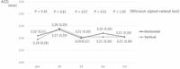
The results were striking. Consistently, the vault measurements from the vertical fixation group were approximately 150 micrometers lower than those of the horizontal group at all observed postoperative intervals. At the one-month follow-up, the vault values stood at 549.0 micrometers in the horizontal group versus 365.0 micrometers in the vertical group. This substantial difference raises implications for patient selection and surgical decisions in myopia correction.
"Vertical ICL fixation reduced the vault by approximately 150 μm compared to horizontal fixation, a deviation from preoperative predictions, which should be considered when determining ICL size," the authors highlighted. Understanding these variances in postoperative vault can guide surgeons in selecting the proper ICL sizes, ultimately enhancing patient outcomes.
The methodology employed was robust, utilizing a prospective case series design with thorough biometric measurements taken at several key postoperative time points: 2 hours, 1 day, 1 week, and 1 month. Throughout the study, researchers meticulously tracked data, ensuring the absence of adverse effects and verifying that anterior chamber depth (ACD) remained stable across both groups without significant differences.
Background context is critical as this study challenges the prevailing norms in ICL surgeries. Traditionally, horizontal fixation is favored based on FDA recommendations, which advocate for techniques that minimize risks to the central cornea while providing adequate astigmatism correction. However, given that many patients present with with-the-rule astigmatism, which can be effectively treated via superior incisions, an increase in vertical fixation surgeries has been noted.
Despite the clear advantages of horizontal fixation, particularly in aligning with existing surgical guidelines and the anatomical characteristics of the eye, this study underscores the potential drawbacks inherent in vertical fixation. As vault measurements are vital for ensuring proper lens positioning and minimizing postoperative complications, the findings present compelling evidence for practitioners to rethink their approaches based on surgical orientation.
The implications extend beyond immediate surgery outcomes. The authors attribute the vault differences to the anatomical variations in positioning within the ciliary sulcus, which may be enhanced when ICLs are fixed vertically, particularly for lenses designed with horizontal fixation in mind. The difference in predicted versus actual vault measurements—where the vertical group averaged a discrepancy of -154.0 micrometers from preoperative predictions—further emphasizes the need for advanced biometric tools and customized surgical strategies.
Moving forward, the need for refined biometric methods cannot be overstated. Surgeons might leverage results from this study to develop size determination nomograms tailored for vertically fixed ICLs, ensuring that similar discrepancies are accounted for. The authors suggest that improved methodologies, such as direct measurements of sulcus dimensions and a deeper understanding of anatomical differences, should be prioritized in future studies to enhance the accuracy of ICL placements.
This research contributes a vital piece to the ongoing discussion regarding ICL surgery and fixation techniques. By illuminating the need for precision in surgical practices, the findings advocate for a collaborative approach among surgeons to continually refine techniques based on empirical evidence. As these studies unfold, they offer promising pathways to personalized and effective myopia correction.
The study stands as a testament to the precision and adaptability required in contemporary ophthalmological practices, as the field progresses toward achieving optimal patient-centric outcomes. The quest for perfecting myopia interventions is ongoing, and each discovery, including the nuances of fixation orientation, brings us closer to shaping clearer visions for millions.