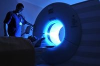
In a groundbreaking study, researchers have revealed that abdominal CT muscle metrics taken from imaging of healthy young individuals could significantly enhance the opportunistic screening for sarcopenia and improve clinical risk stratification. This research, led by Dr. Connie Ju of Stanford University, was published on May 7, 2025, in the American Journal of Roentgenology. The findings provide reference mean values and standardized cutoffs analogous to a T-score of -2 for skeletal muscle index and skeletal muscle density at the L3 level on abdominal CT, potentially aiding in early identification of sarcopenia.
Skeletal muscle health is crucial as it serves as a "vital positive bellwether of overall health," with loss of normal muscle mass indicating increased risk for diseases such as cardiovascular conditions, cancer, or diabetes. Early identification of sarcopenia allows clinicians to initiate timely interventions with patients. However, the researchers noted that "reference values applicable across broad populations are lacking," which prompted their study.
To address this gap, Ju and her colleagues conducted a comprehensive literature review estimating reference cutoff values for CT skeletal muscle metrics. The team analyzed 14 studies that reported skeletal muscle index (SMI) and/or skeletal muscle density (SMD) on CT at the L3 vertebral level in healthy young adults aged 18 to 45. This review involved data from 16,958 individuals, including 11,819 men and 5,139 women, tracking SMI and SMD information.
The results were striking: the estimated global mean value for skeletal muscle index was found to be 54.6 in men and 42.4 in women. In terms of skeletal muscle density, the mean was reported as 47.4 HU in men and 43.6 HU in women. The cutoff values corresponding with a bone health T-score of -2 were determined to be 36.3 in men and 27.5 in women for skeletal muscle index, while for skeletal muscle density, the values were 36.4 HU in men and 28.1 HU in women.
Future research could explore whether using these two metrics in combination is clinically advantageous compared to using just one. The authors also noted ongoing research evaluating the potential benefits of a multivariable sarcopenia Z-score, akin to the Z-score used in dual-energy x-ray absorptiometry (DXA) for osteoporosis, integrating CT muscle metrics with patient age and other important variables such as body mass index or CT-based adiposity metrics.
In another innovative study published in the Lancet Digital Health, researchers from Mass General Brigham introduced a new AI tool called FaceAge, which uses selfies to estimate a person's biological age. This tool could assist doctors in evaluating how well patients might respond to cancer treatment. The concept revolves around the idea that humans age at different rates, and physical appearance can provide insight into a person's "biological age"—a measure of their physiological age, which is influenced by lifestyle and genetics.
Dr. Hugo Aerts, the corresponding author of the study, emphasized the significance of this development, stating, "Our study now has shown for the first time that we can really use AI to turn a selfie into a real biomarker source of ageing." The algorithm was trained using 59,000 photos, demonstrating its potential to provide a low-cost, repeatable method to track an individual’s biological age over time.
The study involved 6,200 cancer patients and found that their biological age was, on average, five years older than their chronological age. The researchers also discovered that older FaceAge readings were linked to worse survival outcomes, particularly in patients with a FaceAge older than 85 years. Dr. Ray Mak, co-senior author of the paper, noted that AI could serve as an objective measure of biological age, complementing traditional vital signs and lab results.
Despite the promising applications of FaceAge, the researchers stressed that AI tools like this should assist clinical decision-making rather than replace clinician judgment. Further studies are underway to explore the tool's applicability to other conditions and the potential impact of cosmetic procedures on its accuracy.
On a different front, the U.S. Food and Drug Administration (FDA) is taking steps to phase out animal testing in the development of monoclonal antibodies and other drugs. The FDA's initiative aligns with the rise of precision medicine and the development of non-animal preclinical testing tools that mimic human biology. This policy shift, supported by the bipartisan FDA Modernization Act 2.0, allows drugmakers to submit non-animal preclinical data when seeking FDA approval for human clinical trials.
On April 10, 2025, the FDA presented a roadmap for implementing this policy over the next three years, aiming to make animal safety and toxicity testing the exception rather than the rule. FDA Commissioner Martin Makary stated, "For too long, drug manufacturers have performed additional animal testing of drugs that have data in broad human use internationally." This initiative is anticipated to accelerate the development of effective treatments while reducing the reliance on animal testing.
Experts in the field, such as Scott Younger from Children's Mercy Research Institute, have expressed caution regarding the complete elimination of animal testing, emphasizing the need for thoughtful implementation of new technologies. The FDA's policy has been met with enthusiasm from companies developing alternatives, such as organoids and organs-on-a-chip, which offer more accurate reflections of human biology compared to traditional models.
Despite the optimism surrounding these advancements, critics argue that the science behind new methodologies is not yet robust enough to fully replace animal testing. Matthew Bailey from the National Association for Biomedical Research highlighted that many reasons contribute to drug failures, not solely the use of animal models. The FDA's announcement has prompted a reevaluation of the role of animal testing in drug development, with calls for a balanced approach that incorporates both traditional and innovative methods.
The FDA's shift represents a significant turning point in drug evaluation, with the potential to enhance the success rates of clinical trials and reduce the costs associated with failed oncology trials, which have reached an estimated $50 billion to $60 billion across the industry. Supporters of alternative preclinical technologies believe that human-derived 3D cell models could improve the chances of drugs successfully reaching the market, addressing the urgent need for effective therapies.
As the landscape of medical research continues to evolve, the integration of new technologies, such as AI and advanced biological models, offers hope for more personalized and effective treatments for patients. The convergence of these innovations marks a promising future for healthcare, with the potential to significantly enhance patient outcomes and the efficiency of drug development.