In a promising advancement for oral cancer detection, researchers have developed a portable fluorescence device that integrates advanced artificial intelligence (AI) technology to identify mucosal lesions at an early stage. This innovative tool aims to address the pressing need for non-invasive methods to diagnose conditions such as oral squamous cell carcinoma (OSCC) and dysplasia, which can often progress unnoticed until they reach late stages.
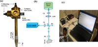
The newly designed device operates using a 405 nm laser diode that emits light onto the oral cavity. This process stimulates two endogenous fluorophores within the tissue—flavin adenine dinucleotide (FAD) and porphyrin. Lead researcher Dr. Pavan Kumar noted, “We have also contributed towards it by developing a steady-state fluorescence-based portable device.” Researchers observed an increase in porphyrin fluorescence specifically in patients diagnosed with cancer, thereby enhancing its detection capabilities.
Traditional methods for diagnosing oral lesions often involve invasive procedures such as biopsies, which can lead to patient discomfort and delay crucial treatment. The introduction of this device provides an alternative that not only minimizes discomfort but also allows for quicker diagnostic responses. The research responds to a critical shortfall; patients diagnosed with oral cancer historically have faced low survival rates—approximately 45% over five years—largely due to late diagnoses.
Utilizing this device, scientists were able to perform examinations on 79 buccal mucosal sites from 36 patients with OSCC, alongside 48 sites from 19 dysplastic patients and 62 control subjects. The fluorescence spectra recorded from these sites highlighted significant differences in the intensity of FAD and porphyrin bands. In particular, a notable 85% of OSCC patients presented detectable porphyrin bands, reinforcing the concept that altered fluorescence profiles could be a reliable indicator of malignant changes in the tissue.
In order to classify and interpret the vast data gathered, the researchers employed AI-based analysis methods including Naïve Bayes, Linear Discriminant Analysis (LDA), and Quadratic Discriminant Analysis (QDA). These methods significantly outperformed standard diagnostic techniques delivering accuracies of 95.34% for OSCC detection and a perfect 100% accuracy for distinguishing between healthy and dysplastic tissues. The authors wrote, "Results manifest that portable device along with the PCA-based QDA tool could be a good substitute for in-vivo identification of oral mucosal lesions." This highlights the device’s potential to not only detect but also to classify various stages of oral cancers accurately.
Moreover, the portability of the device makes it especially advantageous for clinical use in diverse settings, ensuring that healthcare providers can utilize it efficiently while catering to patient needs. The team conducted tests in the Hallet Hospital (Lala Lajpat Rai Hospital) in Kanpur, India, confirming the feasibility of the device for real-time diagnostics in clinical environments.
As part of the study, ethical approval was obtained from the Indian Clinical Trials Registry, illustrating the commitment to maintaining healthcare standards while advancing diagnostic technologies. The results reveal an urgent need for effective early detection methods, reinforcing the role of technology in improving patient outcomes.
Looking ahead, the researchers are hopeful that this device can seamlessly integrate into regular screening processes, potentially revolutionizing the way oral cancers are diagnosed and managed. With continuous advancements in AI and optical technologies, the future is bright for developing tools that can make early detection of malignancies not only possible but also more accessible.
The study’s findings pave the way for future investigations into the use of similar devices in other forms of cancer detection, offering a template for innovation in medical diagnostics aimed at saving lives.