A novel MRI-based tool, segfatMR, has shown promise in accurately quantifying abdominal fat compartments, a finding significant in obesity research and related health metrics. This semiautomatic segmentation software significantly outperforms traditional methods, promising to be a decisive asset in clinical practices and research settings.
Recent research highlights the increasing role of abdominal adipose tissue (AT) as biomarkers for diseases like type 2 diabetes, cardiovascular disease, and cancer. Obesity, a growing epidemic, presents serious health implications, and effective management necessitates accurate measurement tools. Traditional imaging methods such as CT and MRI serve essential roles; however, MRI is preferred due to its lack of ionizing radiation.
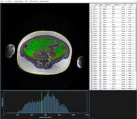
The innovative segfatMR tool was developed through a collaboration at the Integrated Research and Treatment Center AdiosityDiseases in Leipzig, Germany. Validation involved a retrospective analysis of MRI datasets from 20 patients aged 25 to 63 years, reflecting a range of body mass indices from 28.3 to 58.8 kg/m². Two independent expert readers conducted the analysis using both segfatMR and the established commercial tool, sliceOmatic, focusing on subcutaneous adipose tissue (SAT) and visceral adipose tissue (VAT).
Results indicated that segfatMR demonstrated excellent agreement with sliceOmatic, with coefficients of determination (R²) exceeding 0.99 for SAT segmentation and around 0.90 for VAT. Moreover, the new tool was nearly twice as fast, with times measuring approximately 25 seconds per slice compared to 40 seconds with sliceOmatic.
The enhancement of speed in analyzing abdominopelvic data could have considerable implications for clinicians and researchers. The overall efficiency can facilitate longer studies and potentially allow for more extensive patient data management without an increase in workload. As stated by the authors of the article, the presented tool “enables a fast and accurate quantification of whole abdominopelvic adipose tissue volume in obesity studies.”
Furthermore, the open-source framework of segfatMR means it can adapt and evolve alongside new imaging techniques, giving it lasting relevance in the rapidly advancing field of medical imaging.
As obesity rates continue to rise globally, the need for tools like segfatMR becomes pressing. With health risks associated with excessive visceral fat being particularly serious, reliable assessments become crucial. The variability in fat distribution and its impact on metabolic health underscores the necessity for precise measurement tools that can capture the nuances of individual anatomical differences.
The validation study details a process where MRI images of the abdominal region were analyzed following a robust methodology. Each patient underwent imaging on a Philips Achieva XR 1.5T MRI system, and image data were processed on standard computing hardware.
Artificial intelligence and deep learning progression is paving the way for fully automated solutions, but current manual intervention remains a critical component, especially in studies involving diverse cohorts with variable fat distribution. SegfatMR serves as an adaptive tool allowing for visual inspection whilst offering the speed of semi-automation.
Despite its advantages, there are limitations to study outcomes—namely, the reliance on specific imaging protocol and the challenge of ensuring consistency across diverse patient populations. However, the tool’s low computational requirements ensure accessibility in less equipped research centers, broadening its potential usage.
As advancements in imaging technologies develop, integrating techniques such as multi-echo Dixon MRI may enhance the reliability and applicability of fat quantification strategies further. The authors see the significance of combining emerging methodologies, stating, “Segmentation tasks will likely benefit from the ongoing progress in deep learning techniques.”
In conclusion, segfatMR represents a promising step forward in the quest for efficient, precise adipose tissue segmentation, namely within the context of rising obesity rates that necessitate effective monitoring and intervention strategies. Future applications of this tool are expected to grow alongside medical research efforts that continuously aim to improve disease management through precise measurement techniques.
This innovative segmentation tool notably embodies a blend of speed and accuracy, which could prove decisive in addressing the implications of obesity for public health.