Researchers at the University of Louisville have developed a novel mouse model of proliferative vitreoretinopathy (PVR), a condition that complicates about 5–10% of retinal detachment cases, leading to surgical intervention failures. This groundbreaking model utilizes a dispase treatment that simulates ocular trauma, enabling scientists to explore the pivotal role played by retinal pigment epithelial (RPE) cells in PVR pathology.
PVR is a serious complication characterized by the growth of fibrous membranes on or within the retina, which is largely attributed to cellular responses following retinal damage from trauma or surgery. Current animal models, which often rely on external cell inputs or synthetic constructs, do not accurately replicate the complex biological processes that occur in human eyes.
In their study published on March 21, 2025, the team sought to address these limitations by establishing a reliable RPE-tagged mouse PVR model. "We developed an RPE-tagged mouse PVR model that recapitulates features of human PVR, allowing us to investigate the roles of RPE cells in the pathology," wrote the authors of the article, illustrating their innovative approach.
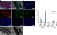
The researchers used C57BL/6 and BALB/c mice, treating them with dispase to damage the retina, which mimicked the conditions leading to PVR. Remarkably, the immunostaining of patient epiretinal membranes confirmed the participation of RPE cells in PVR development. This was evident as 10 out of 14 patient membranes showed pigmentation, indicating a significant RPE presence. Furthermore, quantitative PCR analysis conducted on RPE showed a dedifferentiation from an epithelial state to a mesenchymal one, crucially switching the cells into a proliferative, mobile state underlying PVR pathology. "Quantitative PCR results indicate the dedifferentiation of RPE cells switches their phenotype to a proliferative and fibrotic state underlying PVR," noted the authors. These findings set the stage for deeper investigations into the molecular mechanisms behind RPE behavior during PVR onset.
In establishing their model, the researchers approached PVR induction using multiple methods, including controlled intravitreal injections of dispase. Mice injected with dispase underwent notable changes, with 100% of treated C57BL/6 mice developing PVR-like characteristics within seven weeks. In contrast, subjecting some eyes to a combined dispase and manual retinal massage yielded lower PVR development rates, indicating the need for carefully calibrated treatment protocols.
Additionally, dasatinib, an inhibitor known to target cellular pathways involved in fibrosis, was examined for its therapeutic benefits. The study discovered that administration of dasatinib effectively reduced the dispase-induced PVR-like phenotype in mice, showcasing its potential for future therapeutic strategies.
The researchers carefully documented the migration of RPE cells post-injury, observing that some cells detached from the RPE layer and formed fibrotic membranes encapsulating the retina. This migration aligns with the understanding of RPE cells gaining myofibroblast characteristics, allowing them to contribute to membrane formation—a significant factor in PVR's pathogenesis.
To validate their findings on genetic expressions, the study incorporated microarray data sourced from the National Center for Biotechnology Information (NCBI). A comparison revealed parallels between gene expression profiles from human PVR retinas and the mouse PVR model, solidifying the model's relevance and utility for future explorations into the condition.
In summary, the developments from the University of Louisville not only enhance our understanding of PVR but also open new avenues for research into targeted therapies. The RPE-tagged mouse model promises to be an essential tool for studying the underlying mechanisms of PVR and identifying potential treatment modalities to mitigate this debilitating condition.