Recent advancements in atomic force microscopy (AFM) have led to the development of a new platform for analyzing single nonadherent cells, a significant step for drug discovery and cellular mechanics studies. Researchers have created a reusable poly(dimethylsiloxane)-based (PDMS) microwell array with micron-sized traps designed to facilitate high-throughput analysis of cells. This innovative system integrates cutting-edge deep learning models for automated data analysis, resulting in more efficient and accurate measurements of cell deformability.
The study, published on March 20, 2025, addresses the challenges faced in traditional AFM practices, particularly when working with nonadherent cells, which often slide away during measurements. The newly designed microwell array traps cells securely in place, minimizing movement and allowing for precise mechanical analysis using AFM compression. This design improves the workflow in cellular assays, allowing researchers to employ AFM to obtain vital insights into cellular mechanics and drug interactions.
Using the microwell platform, researchers examined Jurkat T-cells, a type of immune cell commonly used in biomedical research. These cells were treated with various cytoskeletal drugs—Y-27632, Blebbistatin, and Cytochlasin B—before AFM measurements. Results showed the cytoskeletal drugs induced significant changes in cellular elasticity. Specifically, Y-27632 and Blebbistatin resulted in decreases of 36% and 47% in Young’s modulus, respectively, indicating a reduction in cell stiffness under these treatments.
This study reveals a groundbreaking application of deep learning techniques in analyzing AFM data. The researchers integrated a custom convolutional neural network (CNN) architecture that excelled at predicting the Young’s modulus from raw AFM data. Their CNN model achieved a coefficient of determination of 0.47 for elasticity predictions and exhibited accuracy scores exceeding 90% in drug classification tasks, including binary detection of drug presence and identification of drug types.
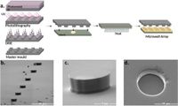
The microwell array fabrication involves a simple and rapid process using photolithographically patterned silicon molds, which allows for high reproducibility and uniformity across multiple PDMS arrays. With significant potential for drug detection applications, this method can facilitate high-throughput screening necessary for drug discovery, making analysis faster and more reliable.
Moreover, utilizing nested leave-one-sample-out cross-validation approaches, the deep learning classifiers demonstrated robust predictive abilities. With average area under the curve (AUC) scores of 0.91 in binary classification scenarios, the models provide a promising framework for future biomedical applications, particularly in automating cellular mechanical property assessments and drug testing processes.
This advancement not only enhances existing AFM techniques but can also be adapted for various cell types and drug investigations. The design allows researchers to fine-tune the microwell sizes for specific cell types while employing machine learning algorithms to extract key parameters quickly and accurately.
In conclusion, the authors emphasize that this innovative platform significantly streamlines the complexity of AFM analysis, paving the way for more efficient high-throughput drug screening and cellular research. The integration of machine learning into this methodology promises transformative impacts on how researchers study cellular mechanics and drug interactions, potentially leading to groundbreaking discoveries in various fields of medical science and pharmacology.