In the realm of endocrine surgery, precise localization of parathyroid adenomas stands as a critical step for successful intervention in patients suffering from primary hyperparathyroidism. A new study published on March 23, 2025, underscores the efficacy of 18 F-fluorocholine PET-CT as a compelling alternative to conventional imaging techniques for preoperative localization of these adenomas.
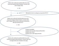
Conducted at Brest University Hospital, this retrospective study involved 156 patients who underwent parathyroidectomy between 2017 and 2023. The majority of the participants (78%) were women, with an average age of 63 years. Primary hyperparathyroidism, characterized by excessive production of parathyroid hormone—often due to a benign tumor known as a parathyroid adenoma—can lead to severe complications. Surgery remains the definitive treatment, making preoperative localization essential.
Current imaging modalities, including cervical ultrasound and [99mTc]-MIBI scintigraphy, have been standard; however, their sensitivity varied significantly. In the study, cervical ultrasound demonstrated a sensitivity of only 60.14%, while [99mTc]-MIBI scintigraphy fared even worse at 46.21%. In contrast, 18 F-fluorocholine PET-CT emerged with an impressive sensitivity of 95.97%, achieving 100% effectiveness for identifying multiple adenomas—a situation where traditional methods often falter.
The authors noted, "18 F-fluorocholine PET-CT is a valuable imaging modality for the preoperative localisation of parathyroid adenomas in primary hyperparathyroidism, with superior performance compared to conventional imaging modalities." This assertion is corroborated by findings that showed the PET-CT method significantly outperformed both cervical ultrasound and scintigraphy, particularly in cases where initial imaging had yielded inconclusive results.
The study also highlighted patient demographics relating to imaging performance. Notably, men showed better results with cervical ultrasound (p = 0.005), while larger adenomas were better detected using MIBI scintigraphy (p = 0.026). Perhaps most critical for clinical practice, elevated PTH levels negatively impacted the performance of 18 F-fluorocholine PET-CT (p = 0.023), indicating that further investigation is warranted for patients with high PTH levels prior to using this imaging method.
As a relatively modern imaging technique, 18 F-fluorocholine PET-CT's adoption signifies a shift towards more accurate diagnostic capabilities in endocrine surgery. With the combined sensitivity of ultrasound and scintigraphy achieving only 80.1%, the new PET-CT technology's rates encourage a reevaluation of its role in diagnosing parathyroid conditions.
While the study underscores promising advancements, doctors must also be cautious regarding the economic implications of utilizing 18 F-fluorocholine PET-CT. Costing approximately €881 compared to €268 for scintigraphy and €37 for standard ultrasound, access may be limited in certain geographical areas.
In summary, the findings from Brest University Hospital significantly bolster the case for implementing 18 F-fluorocholine PET-CT as a first-line imaging modality in clinical settings, especially for patients exhibiting thyroid nodules or confirmed multigland disease where traditional methods have historically fallen short. As surgical techniques and imaging modalities advance, patients suffering from primary hyperparathyroidism may benefit from these innovations, paving the way for better outcomes and more refined surgical interventions.