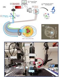
A novel lab-in-a-fiber (LiF) device has emerged as a promising solution for cancer diagnostics by enabling the selective detection and collection of single cells or small clusters. This groundbreaking technology combines an optical fiber with a fiber capillary, allowing researchers to capture fluorescently labeled tumor cells from a mixture of unlabeled cells, overcoming challenges of traditional biopsies and liquid biopsies.
Developed by a team of scientists, the LiF device demonstrates potential applications in both in vitro and in vivo diagnostics, promising less invasive methods for cell sampling and analysis. This innovative approach not only reduces the costs associated with cancer diagnostics but also simplifies the process by making it compatible with standard laboratory equipment.
The LiF device's capillary system employs a multimode silica fiber (MMF) and a fiber capillary with precise dimensions, enabling the accurate selection of target cells based on their fluorescence. The system operates by illuminating the sample with a blue laser, thus exciting fluorescent cells, which then light up for detection. A negative pressure mechanism allows for effective collection by sucking selected cells into the capillary.
One of the challenges faced in fluorescence detection is photobleaching, which limits the measurement time of labeled cells. The LiF device employs a novel pulsed excitation technique, illuminating fluorescent dyes intermittently to extend the usage time significantly. Traditional continuous excitation methods lead to rapid decay of fluorescence, with signal halving in just over 130 milliseconds. By switching to a pulsed system operating at 100 Hz, the team achieved a much slower decay, thus increasing the efficiency of detection.
The experiments conducted using cancer cells, specifically MCF-7 cells, showed about 90% viability of captured cells post-collection, ensuring that the cells retained their essential properties for further analysis. This high level of cell viability marks a significant advancement over conventional approaches, where samples often suffer from deterioration during collection procedures.
Details described in the study indicate that functional capabilities of the LiF system allow it to scan through large volumes of fluid, detecting changes in cell concentrations over time, which could be important for monitoring tumor progression and treatment efficacy. Future aspirations for the technology include integrating artificial intelligence to further enhance diagnostic accuracy using data derived from the patterned fluorescence intensities of captured cells.
The potential applications of the LiF device are vast. As research gears towards minimizing invasiveness in medical procedures, its compact size and flexibility make it an ideal candidate for remote and sensitive regions where traditional biopsy might be more difficult or risky.
The delivery of such technology could pave the way for new personalized methods to treat cancer, wherein molecular characteristics of each patient's tumor type can be identified with precision. By using this innovative fiber-optic approach, the future of cancer diagnostics looks promising, emphasizing a pathway towards more accessible and less invasive testing methods.
This combination of optical fiber technology with advanced cell detection processes marks a notable step forward in medical diagnostics, aligning with the increased need for minimally invasive procedures across various fields, particularly in oncology.