In a groundbreaking advancement in medical imaging, researchers have introduced a new groupwise multiresolution network poised to enhance the accuracy of image registration in dynamic contrast-enhanced magnetic resonance imaging (DCE-MRI), especially in assessing lung perfusion in pediatric patients with congenital diaphragmatic hernia (CDH). The method cleverly combines traditional image registration techniques with the speed and efficiency of deep learning algorithms.
The study, published in Scientific Reports, aims to overcome the challenges posed by patient movement during scans, which can lead to inaccuracies in visualizing organ function. The proposed network utilizes multiple resolutions and is trained through unsupervised learning, significantly improving the spatial alignment of images while reducing processing time.
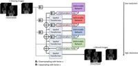
The backdrop for this scientific leap lies in the notorious difficulty of registering images—aligning different scans to present a unified picture, especially when examining temporal changes in anatomy caused by breathing or other movements. Existing methods employed pairwise registration strategies that rely on selecting a fixed reference image, a process that can introduce significant bias and inaccuracies.
To tackle this issue, the researchers, led by Anna Strittmatter, leveraged a dataset comprising 30 DCE-MR scans of 2-year-old patients who had undergone repair for CDH. Each scan was performed using a high-resolution three-dimensional TWIST sequence at a 3T MRI scanner, resulting in 50 time steps measured over a temporal period of 1.5 seconds each. “In the context of our experiments, using a groupwise approach allowed us to avoid the pitfalls associated with fixed-image selection,” said the authors of the article. “By averaging the images to create a template, we improved the alignment process.”
The methodology involved training the network for 200 epochs on NVIDIA A100 GPUs, using advanced loss functions to fine-tune the image transformations. This approach allowed for rapid registration of the four-dimensional scans, achieving processing times under 10 seconds—a substantial improvement over traditional methods like SimpleElastix, which can take over 14 minutes.
Evaluation metric calculations yielded impressive results, with the new groupwise multiresolution network recording a Structural Similarity Index (SSIM) of up to 0.953, indicating high spatial alignment. This level of precision is crucial for generating accurate lung perfusion maps, essential for evaluating patients with CDH.
As the authors noted, “Image registration with our proposed groupwise network enhances the accuracy of medical image analysis by leading to more homogeneous perfusion maps.” On the clinical side, this improvement could translate into better patient outcomes through enhanced diagnostics and treatment planning.
The registration technique not only demonstrated enhanced image quality but also alleviated issues related to medically implausible transformations seen in many classical approaches. The performance of their method outstripped both pairwise and established groupwise registration techniques, affirming the efficacy of deep learning applications in the realm of medical imaging.
Future research directions indicated by the authors include applying this methodology across diverse medical imaging realms and potentially refining the algorithms for even greater efficiency, precision, and applicability beyond pediatric contexts. As the field of medical imaging continues to evolve, this new groupwise multiresolution network represents a significant step forward, paving the way for future innovations.
The authors maintain that deploying machine learning techniques such as this in medical imaging not only fosters greater precision but also assists clinicians in harnessing the full potential of imaging data for improved patient care.