A groundbreaking advancement in skin research has taken a step forward with the development of a microneedle-based culture technique, allowing for sustained live imaging of excised human skin tissue. This innovative method not only preserves the skin tissue’s viability for extended periods but also enables researchers to observe cellular behavior and tissue dynamics at an unprecedented level of detail.
Traditionally, live imaging of human skin has been limited due to the invasive nature of existing methods, which often require surgical exposure and anesthesia. Aiming to address these limitations, researchers devised a non-invasive approach that utilizes microneedles to maintain the skin tissue in an environment mimicking that of a living organism. This research, led by Tohgasaki et al., was published in Scientific Reports and provides new insights into skin metabolism, healing processes, and reactions to various stimuli.
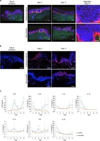
The microneedle technique involved cultured excised human skin samples, integrating a method to inject culture medium directly into the tissue. Unlike conventional methods that often resulted in cell death and separation of the epidermis and dermis after just a few days, the microneedle approach showed significant improvements. The researchers observed that after 5 days, the tissue retained its structure and cellular integrity, allowing for the long-term observation of keratinocytes, collagen, elastin, and other essential cellular components.
Key findings from the study revealed that traditional culture methods caused substantial cellular damage. For instance, epidermal cells from samples cultured using conventional techniques displayed smaller, nearly perfectly circular nuclei, indicative of deterioration. In contrast, tissues maintained with microneedles exhibited almost no DNA damage and preserved their structural integrity, remaining bonded across the epidermal-dermal junction after 11 days.
Significantly, levels of lactate dehydrogenase (LDH) and various inflammatory cytokines (including IL-6, IL-10, and TNF) were considerably lower in the microneedle-cultured tissues compared to those subjected to traditional methods. This reduction in cytokines points to a decreased inflammatory response, suggesting enhanced viability and functionality of the tissue.
Furthermore, the research successfully captured time-lapse imaging of critical skin appendages and fatty tissues, allowing for a dynamic observation of capillaries, hair follicles, and even lipid-laden adipocytes. The investigators utilized fluorescent probes to analyze cellular components such as nuclei, mitochondria, and reactive oxygen species (ROS), which were visualized throughout the time-lapse observations.
Among the most compelling observations, the researchers noted migratory activities within the matrix at the epidermal-dermal junction, revealing interactions and movements that had previously been unobserved in excised human tissue. High-resolution imaging allowed for quantification of membrane movements and dynamic changes that support our understanding of skin physiology.
The implications of this study are vast. The potential to conduct long-term live imaging of human skin could enhance dermatological research significantly, allowing scientists to explore aging, chronic inflammation, and wound healing phenomena in real-time. Moreover, the microneedle approach shows promise in minimizing invasiveness for various medical applications, including painless drug delivery and testing drug responses in cultured human skin.
The researchers caution, however, that while the microneedle technique shows great promise, variability in human tissue and the necessity for longer-term evaluations remain as areas needing further study. The novel culture method introduced not only enhances our comprehension of skin biology and dynamics but also paves the way for future developments in dermatological treatments.
In conclusion, this innovative microneedle culture technique represents a significant leap in non-invasive skin research, facilitating extensive exploration of human tissue functionalities and potentially informing novel therapeutic strategies.