Recent research has shed light on the intricacies of skin microcirculation, specifically focusing on the structural differences and distribution patterns of dermal microarterioles and microvenules in human forehead skin. Published in Scientific Reports, this study utilized an innovative intravascular dual perfusion technique combined with immunofluorescence staining to provide a clearer picture of the vascular architecture in this critical area.
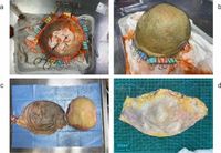
Understanding skin microcirculation is paramount in clinical practice. It plays a vital role in revealing the vascular mechanisms underlying systemic diseases and is essential for successful skin surgeries, particularly flap surgeries where blood flow and recovery are dependent on the density and distribution of microvessels. The research was based on two post-mortem cadaver specimens, where lead oxide-gelatin perfusion was employed to label microarterioles, while latex was used for microvenules.
The results unveiled significant structural differences between dermal layers: vessels situated in the deep dermis exhibited larger diameters and thicker walls than those in the superficial layer, correlating with the functional demands imposed by each dermal layer. Microarteriole diameter measured 23.78 ± 3.88 μm in the superficial dermis and 61.48 ± 23.69 μm in the deep dermis, whereas microvenule diameter was recorded at 40.85 ± 10.61 μm and 87.58 ± 19.27 μm, respectively. This discrepancy in vessel dimensions provides insight into how vessel structure adapts to different physiological conditions.
In addition to diameter, wall thickness was also examined. The wall thickness of microarterioles in the superficial dermis was found to be 3.83 ± 0.49 μm compared to 25.03 ± 6.66 μm in the deep dermis. Similarly, microvenules demonstrated a wall thickness of 2.69 ± 0.45 μm in the superficial dermis, increasing to 5.04 ± 0.67 μm in the deeper layer. These variations illuminate the mechanical demands faced by the vascular components within the skin, where the deeper layers necessitate more robust vessel walls to sustain higher pressures during circulation.
The study also highlighted notable differences in microvessel density. While the superficial dermis contained a higher microarteriole density of 1.96 ± 0.57 vessels/mm² compared to 1.71 ± 0.42 vessels/mm² in the deep dermis, microvenule density was also greater in the superficial dermis at 2.88 ± 0.44 vessels/mm², contrasting with 2.29 ± 0.35 vessels/mm² in the deeper layer. Such differences indicate the adaptation of different dermal layers to their functional roles, with increased densities in the superficial layer supporting greater nutrient and oxygen exchange being crucial for the epidermis situated above.
To better distinguish between these microvessels, the researchers identified monocarboxylate transporter 1 (MCT1) as a specific marker for microvenules, while cluster of differentiation 31 (CD31) served as a general vascular marker applicable to both vessel types. The meticulous analysis revealed that MCT1 staining was exclusively positive in a subset of microvenules, underscoring its utility in differentiating microvenules from microarterioles in dermal tissues. This innovation could serve as a pivotal tool in clinical settings for improving the outcomes of surgical procedures and tissue diagnostics related to microcirculation.
These findings not only advance our understanding of the microcirculatory architecture of the forehead skin but also pave the way for enhancing surgical interventions that rely heavily on these small blood vessels. Effective surgical planning and postoperative care can significantly benefit from an in-depth comprehension of microvessel distribution and characteristics. The insights drawn from this study may usher in an era of improved outcomes in reconstructive surgeries, optimizing techniques such as flap surgeries that often face substantial challenges related to blood flow and healing.
Moreover, the systematic mapping of microvascular structures through advanced imaging techniques and specific immunofluorescent markers represents a crucial enhancement in surgical precision, potentially resolving longstanding debates surrounding the effectiveness of venous superdrainage and arterial superperfusion strategies in enhancing flap survival.
In conclusion, this research lays significant groundwork for future studies aiming to refine our understanding of skin microcirculation and its critical impact on surgical practices. As ongoing efforts are directed toward advancing imaging techniques, the ability to visualize and differentiate these microvessels more effectively in various anatomical regions could herald a significant leap forward in surgical science.