A recent multi-center study has unveiled a promising deep learning (DL) model that utilizes magnetic resonance imaging (MRI) to accurately predict muscle-invasive bladder cancer (MIBC). Researchers examined data from 559 patients between February 2012 and December 2023, demonstrating the potential for improved diagnostic precision in bladder cancer treatments.
Bladder cancer is the tenth most common cancer diagnosed around the world, with approximately 573,000 new cases reported in 2020. Among these, about 25% are classified as muscle-invasive bladder cancer, which is critical for determining treatment options. Traditional diagnostic tools, such as cystoscopy and computed tomography, often yield inaccurate results, with studies indicating that up to 49% of cases were incorrectly staged.
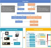
To address this issue, the authors developed an artificial intelligence-based DL model drawing from MRI, aiming to distinguish between MIBC and non-muscle-invasive bladder cancer (NMIBC). Utilizing Inception V3, a robust DL architecture, the model analyzed three-channel image inputs, including original T2-weighted images, segmented bladder images, and regions of interest.
The DL model achieved a remarkable accuracy of 92.4% in the validation set, with a sensitivity of 94.7% and specificity of 91.5%. These figures demonstrate the model's capacity to predict MIBC reliably, offering an enhanced pathway for clinical decision-making.
In further assessments, when tested against an internal cohort, the DL model maintained a 92.1% accuracy, and in an external test set, it recorded an accuracy of 81.6%. However, the sensitivity dropped significantly in the external test, indicating the need for further refinements.
When categorized according to the vesical imaging report and data system (VI-RADS), the DL model exhibited impressive metrics, scoring 93.5% in VI-RADS 2 and 80% in VI-RADS 3, both critical categories for assessing muscle invasion likelihood. The findings suggest that the DL model could serve as a significant complement to existing VI-RADS criteria, enhancing the accuracy of predictions.
The methodology involved a meticulous review of pathological specimens by experienced pathologists, alongside MRI examinations completed under standardized conditions. This systematic approach, combined with advanced imaging technologies, underpins the reliability of the DL model.
Despite the promising outcomes, the study identified challenges associated with the DL model’s performance in classifying NMIBC tumors, especially near the ureteral orifice. Misclassifications tended to occur with tumors in complex locations, prompting the researchers to consider future developments, including potentially using 3D models to improve reliability.
The authors expressed the need for broader training datasets to account for variances across institutions, advocating for multicenter training strategies. Addressing the discrepancies in imaging quality and patient characteristics will be paramount in enhancing the model's robustness and generalizability.
This research demonstrates a significant advancement in employing DL models within medical imaging, specifically for bladder cancer diagnostics. As the technology matures, it holds the promise of revolutionizing how healthcare providers identify muscle invasion in bladder tumors, potentially leading to better patient outcomes.