Brain tumors remain one of the most challenging health issues, with approximately 25,400 new cases expected to be diagnosed in the United States in 2023 alone. Effective diagnosis and treatment depend on precise segmentation of these tumors in magnetic resonance imaging (MRI) scans. A new study proposes an innovative framework that significantly improves the segmentation accuracy of brain tumors using deep learning techniques.
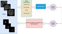
The study introduces a novel segmentation model that integrates a Multiscale Attention U-Net with an EfficientNetB4 encoder to optimize the feature extraction process. EfficientNetB4 is a cutting-edge convolutional neural network that leverages compound scaling to enhance accuracy while minimizing computational overhead. The authors of the article note that "this study demonstrates the effectiveness of the proposed method for robust and efficient segmentation of brain tumors, positioning it as a valuable tool for clinical and research applications.”
Brain lesions pose their immense risk due to the potential for functional impairments. Gliomas, particularly glioblastomas, stand out as some of the most aggressive forms of brain tumors. As highlighted in the article, effective treatment planning requires accurate detection and segmentation of these tumors, which is primarily facilitated through advanced imaging techniques like MRI and computed tomography (CT). However, conventional methods of segmenting tumor areas in MRI images are often labor-intensive and prone to errors, leading to a significant reliance on advanced artificial intelligence (AI) methods.
The proposed framework distinguishes itself by utilizing a Multi-Scale Attention Mechanism, employing convolutional kernels of varying sizes (1×1, 3×3, and 5×5) to enhance feature representation. This approach addresses the limitations of traditional convolutional neural networks (CNNs), which often struggle to capture the nuanced boundaries of tumors. The authors implement attention-enhanced skip connections to improve localization accuracy and minimize irrelevant background noise.
Experimental results demonstrated the model's superiority over other segmentation techniques. On the publicly available Figshare brain tumor dataset, the EfficientNetB4-based model achieved an impressive overall accuracy of 99.79%, with a Dice Coefficient of 0.9339 and an Intersection over Union (IoU) score of 0.8795. Such performance illustrates the potential of incorporating sophisticated neural networks for clinical applications, especially in settings where quick and accurate decision-making is crucial.
In addition to its clinical implications, this research contributes to the ongoing dialogue surrounding deep learning in medical image analysis. Despite substantial advances, challenges remain related to model generalizability and interpretability, especially when considering varied patient populations and imaging protocols. The complexity of integrating such models into clinical workflows also poses significant hurdles. Nonetheless, the study champions a robust path toward improving imaging processes, citing that 2the proposed method strengthens the precision, effectiveness, and reliability of automated medical image analysis.2
Ultimately, the findings from this research signal a pivotal shift in the application of AI in neuroimaging, leveraging technical innovation for real-world medical challenges. As research continues to refine these models and validate their usefulness in diverse clinical settings, the hope is that such advancements will lead to improved outcomes for patients battling brain tumors.