A New Rig Simulates Muscle Function in Lumbar Spine Tests
Researchers develop an advanced setup that enhances in vitro biomechanical studies by mimicking muscle traction.
Numerous research questions demand precise in vitro testing of the lumbar spine and pelvis, particularly concerning biomechanical stability and movement replicability. Traditional testing methods often apply forces through a single vertebral body while fixing another, which has been validated for assessing kinematics and pressures. However, this approach may not accurately represent the complexities of human muscle function. In light of this limitation, researchers have crafted a novel testing rig designed to simulate muscle exertion more accurately, leading to findings that could significantly impact our understanding of spinal biomechanics.
This innovative test rig applies forces directly to the vertebral bodies through artificial muscle attachments, effectively capturing the stabilizing effects of muscles known from existing literature. By placing muscle attachments in close proximity to their anatomical footprints on the bones, the researchers have engineered a system that faithfully mimics the role of human musculature. The project, described in detail by Georg Matziolis and colleagues, utilized linear actuators to simulate three paired muscle groups: the anterior, posterior, and oblique trunk muscles. Coupled with an optical 3D motion capture system from GOM (Zeiss, Germany), the apparatus meticulously measures lumbar spine repositioning against the pelvis during testing.
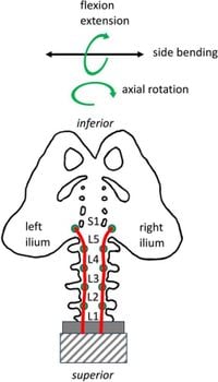
Throughout their experiments, all muscle attachments withstood maximum forces up to 1 kN (kilonewton) without failure. The study recorded a range of motion for the lumbar spine under simulated muscle traction, achieving physiological movements including an extension of 18°, flexion of 27°, lateral bending of 33°, and axial rotation of 11°. Notably, the anterior trunk muscles exhibited the most significant effects during flexion and extension at 0.16 ± 0.06° per newton, while the oblique trunk muscles drove lateral bending and axial rotation with values of 0.17 ± 0.16° and 0.10 ± 0.14°, respectively. The nutation of the sacroiliac joint (SIJ) averaged 1.2° ± 0.2° across the tested cadaveric specimens.
This study, conducted with six cadaveric specimens from T12 to the pelvis, revealed how variances in muscle activity influence lumbar spine movement. The anterior trunk muscles were primarily responsible for flexion, whereas the oblique trunk muscles excelled in lateral movement and rotation. These nuanced insights into muscle interaction potentially reshape how clinicians and researchers approach lumbar spine pathologies and surgical interventions.
Researchers crafted the rig to simulate everyday biomechanical forces seen during various physical activities. With direct attachments of the trunk muscles to vertebral bodies, the researchers can more precisely replicate the forces acting on the spine—a deviation from more conventional models that rely solely on external load application. The transition to this muscle-driven model establishes groundwork for future studies into the dynamic interactions of spinal muscle groups during physical activity.
The experimental protocol underwent thorough ethical scrutiny and was approved by the ethics committee at the Friedrich-Schiller University Jena. Participants provided informed consent while alive to facilitate the use of their cadaveric specimens, ensuring compliance with ethical standards within the scientific community.
The results from this research should have significant implications for both biomechanical testing and clinical outcomes related to spinal health. Addressing the pressures and movements experienced in vivo allows for better alignment with real-world scenarios. Moreover, the apparatus holds potential for adapting to simulate conditions such as sarcopenia or muscular dystrophy, which could lead to remarkable advancements in understanding spinal disorders and improving surgical techniques.
The lumbar spine test rig represents a significant enhancement over existing methods, allowing new paradigms in understanding how different muscle groups contribute to spinal function. The findings of this study not only draw on previous biomechanical models but provide fresh insight into muscle interactions that may be pivotal in preventing or treating conditions related to lumbar instability.
Moving forward, future research may expand the system to incorporate additional muscle groups and further refine the precision with which it mimics in vivo conditions. By delivering comprehensive biomechanical assessments, this rig promises to enhance our understanding of spinal dynamics, illuminating potential pathways for further research and clinical application.